
Easy to miss, challenging to fix: neglected locked posterior shoulder dislocation
BY DR MOSTAFA SAMIR ZIED

Posterior shoulder dislocation is a rare condition that is regularly missed during diagnosis. As a result, the affliction can go unnoticed for weeks, causing patients increasing pain and severely limiting their range of motion. Here, Mostafa Samir Zied outlines the challenges he encountered when operating on a particularly challenging case. He won the World Surgery Tour (WST) 2024 ‘My most challenging case’ competition on myAO in the Sport category with the example case.
I recently came to treat a 22-year-old male patient who presented with pain in his right shoulder that had already persisted for several weeks. The patient also displayed a very limited range of motion in his shoulder: both external rotation and forward flexion were at zero degrees, while abduction was at 30 degrees.
Initially, the patient had presented to an emergency room, where he had stated that his pain was the result of his friend having gently pulled his shoulder while joking around. Upon a subsequent X-ray (fig. 1) the patient had been diagnosed with a shoulder contusion.
However, when his symptoms showed no sign of improvement over the following few weeks, a colleague, in a fresh consultation, diagnosed weakness in the shoulder muscles and advised the patient to start physiotherapy. Again, several weeks passed with no notable improvement.
When the patient ultimately presented to me, I thoroughly examined him, and I re-addressed the question of how he had sustained his injury. He admitted that he had lied about the mode of trauma to the ER staff—in reality, he had sustained direct trauma while playing football, but had been afraid to admit this to his parents.
Subsequent CT and MRI scans provided clarity: the shoulder contusion had been a misdiagnosis. Instead, the patient suffered from a locked posterior shoulder dislocation. The locking occurred between an anterior defect in the humeral head, known as a reverse Hill-Sachs lesion, and the posterior glenoid rim.


Shoulder instability easily leads to misdiagnosis of rare cases
Because it is so highly mobile, the glenohumeral, or shoulder, joint is very susceptible to instability in the form of subluxation or even frank dislocation. However, posterior dislocation of the glenohumeral joint, as exhibited by my patient, is rare: it accounts for less than two percent of all shoulder dislocations.
Posterior shoulder dislocation may occur because of trauma, seizures, or electric shock. However, as it is so rare, misdiagnosis of the injury is very common, occurring in 50 to 79 percent of cases. Especially in ERs, the condition is overlooked regularly. Oftentimes, the physical examination simply is not performed thoroughly enough. Inadequate use of radiographic imaging equipment is another common reason.
In order to be correctly diagnosed, the condition therefore requires a high degree of attentiveness on the part of the treating physician.
The options for surgical treatment of posterior shoulder dislocation include simple reduction or transfer of the subscapularis tendon into the reverse Hill-Sachs lesion, a procedure that has become known as the 'McLaughlin'. If the lesser tuberosity is transferred into the defect along with the subscapularis, it is called the 'modified McLaughlin'. Another option, in cases in which more than 50 percent of the head is damaged, is shoulder arthroplasty.
The surgical procedure in shoulder dislocation
In many dislocations, the shoulder can be repositioned manually via a closed reduction procedure that requires no surgery. However, cases in which the dislocation is neglected for more than three weeks usually require surgical reduction. An unsuccessful trial of closed reduction confirmed that this was true in the case of my 22-year-old patient.
Our preparations for the operation included an in-depth patient briefing during which the procedure and its implications were explained in detail. With the patient under general anesthesia and positioned in the beach chair position, we then initiated the actual operation by draping and scrubbing the right shoulder. Opting for a deltopectoral approach, we made a precise incision in the anterior shoulder region, enabling us to navigate between the pectoralis major and deltoid muscles in order to access the underlying structures.


However, when we reached the humerus, we encountered an unexpected challenge: excessive internal rotation had severely disrupted the normal anatomical order. Through careful dissection, we eventually managed to locate the long head of the biceps, which had concealed itself beneath the conjoined tendon. Recognizing the pivotal role of the long head of the biceps as a gateway to the shoulder joint, we proceeded with the necessary maneuvers to restore normal joint architecture.
Next, we carefully tagged the subscapularis tendon with high tensile sutures. The subscapularis is identifiable by the anterior circumflex humeral artery and two venae comitantes—three vessels that pass at the tendon's lower border and that are commonly known as the 'three sisters'.
In order to gain access to the locked humeral head, we then performed an osteotomy of the lesser tuberosity and its subscapularis attachment and retracted them medially. As a blunt reduction technique, we employed an index finger to form a hook around the humeral neck. This effectively realigned the joint and enabled us to identify the reverse Hill-Sachs lesion affecting the anterior humeral head. The defect was repaired via careful curettage to create a raw area, followed by the insertion of two double-loaded anchors along the periphery.
These anchors were securely sutured to the lesser tuberosity with its subscapularis tendon attachment (modified McLaughlin technique). Additionally, all sutures, including the tagged sutures, were transosseously passed as a lateral row to the bicipital groove. Shoulder stability was assessed intraoperatively in all directions, ensuring optimal function, integrity, and stability of the joint.
Postoperative rehabilitation protocol in shoulder dislocation
The postoperative rehabilitation protocol proceeded as follows:
- For the first four weeks after the procedure, the patient's right shoulder was immobilized in external rotation and internal rotation was not allowed. At the same time, active exercises were prescribed to train the elbow, wrist, and hand.
- After four weeks, physical therapy was initiated. This included exercises to progressively increase passive, active-assistive, and active range of motion, as well as exercises to strengthen the rotator cuff.
- Between eight and twelve weeks after the operation, range restrictions were removed, and exercises to strengthen the muscles continued.
- After the twelfth week full activity was finally allowed.
To summarize, posterior shoulder dislocation is difficult to diagnose. As a result, it is often missed. Part of the reason is that it is so rare—many physicians simply do not think of the condition as a possible cause when patients present to them with shoulder pain. I would therefore urge colleagues to develop a more acute awareness of posterior shoulder dislocation—especially because its negative effects on patients' quality of life can get progressively worse the longer it goes undetected.
About the author

Mostafa Samir Zied, M.D., MSc, EFORT, graduated in 2012 and boasts over 10 years of orthopedic expertise. Formerly associated with Helmya Military Hospital and El-Bank AlAhaly Hospital, he currently serves as a senior registrar at El-Maadi Charity Hospital and Electricity Company Hospital.
He has been practicing sports medicine since 2019.
References and further reading:
- Hatzis N, Kaar TK, Wirth MA, Rockwood CA Jr. The often overlooked posterior dislocation of the shoulder. Tex Med. 2001; 97(11):62-67.
- Kelly JP. Fractures complicating electro-convulsive therapy and chronic epilepsy. J Bone Joint Surg Br. 1954; 36(1):70-79.
- Charalambous CP, Gullett TK, Ravenscroft MJ. A modification of the McLaughlin procedure for persistent shoulder instability: technical note. Arch Orthop Trauma Surg. 2009; 129(6):753-755.
- Kokkalis ZT, Mavrogenis AF, Ballas EG, Papanastasiou J, Papagelopoulos PJ. Modified McLaughlin technique for neglected locked posterior dislocation of the shoulder. Orthopedics. 2013 Jul;36(7):e912-6. doi: 10.3928/01477447-20130624-22.
You might also be interested in:

myAO
Gain access to trusted sources of knowledge, moderated case discussions, and a community of 80,000 verified surgeons worldwide.
AO Recon courses and events
Find all upcoming AO Recon courses, webinars, and online events in your region and worldwide.
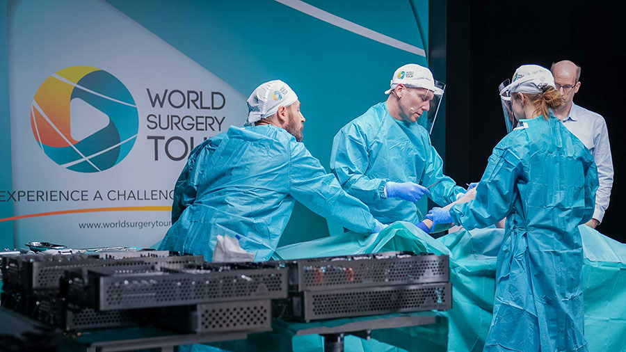
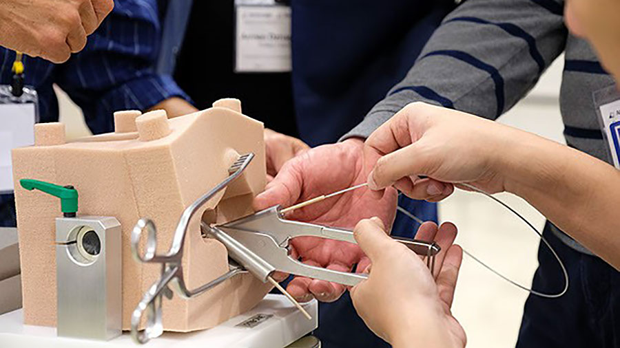
World Surgery Tour (WST)
An innovative education platform for ortho and trauma professionals to unlocks skills, knowledge, and career advancements.